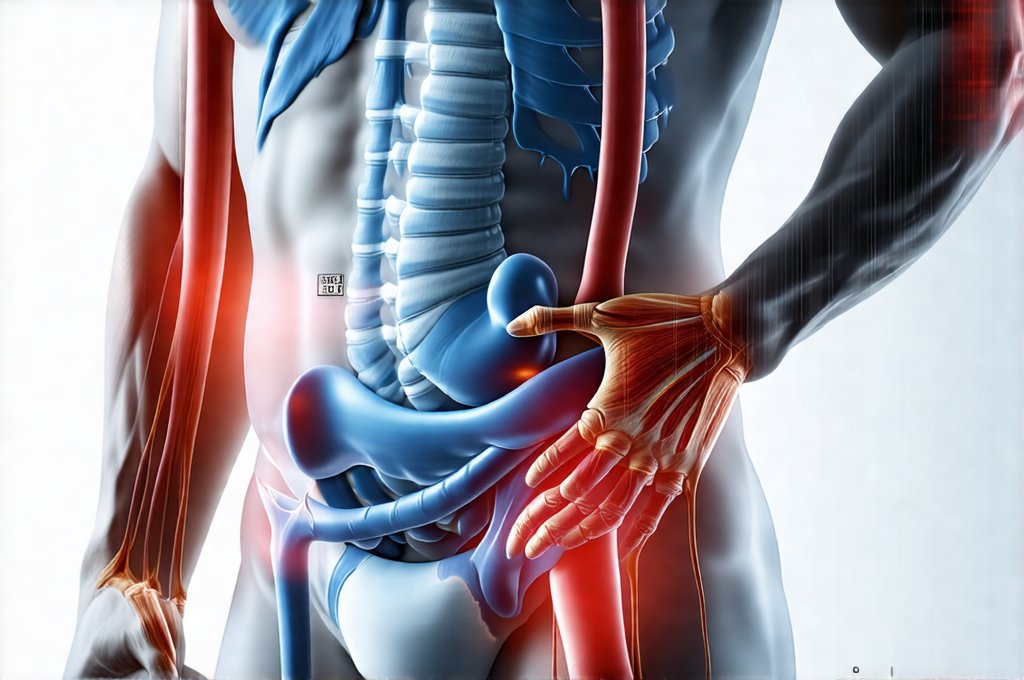
Abdominal pain is arguably one of the most common reasons people seek medical attention. Its broad spectrum of possible causes – ranging from relatively benign indigestion to life-threatening emergencies – makes diagnosis challenging. The location, character (sharp, dull, cramping), timing, and associated symptoms all provide clues, but often imaging and other scans are essential to pinpoint the source of discomfort and guide treatment decisions. Effectively navigating this diagnostic process requires understanding which scans offer the most relevant information for different presentations of abdominal pain, and how those scans complement a thorough clinical evaluation performed by a healthcare professional. This article will explore the various scanning modalities used in diagnosing abdominal pain, outlining their strengths, weaknesses, and typical applications.
The selection of appropriate imaging is not a one-size-fits-all approach. It depends heavily on the patient’s specific symptoms, medical history, physical examination findings, and initial laboratory tests. A physician will consider factors like age, pregnancy status (if applicable), and any pre-existing conditions before ordering scans to minimize radiation exposure and ensure the most accurate diagnosis with the least invasive methods possible. Importantly, imaging is rarely used in isolation; it’s integrated into a broader clinical picture to arrive at a definitive diagnosis and treatment plan. It’s also critical to remember that while scans provide valuable information, they are tools assisting medical professionals – interpreting scan results requires expertise and contextual understanding. If you suspect a food intolerance may be the source of your discomfort, consider identifying food intolerances that cause abdominal pain.
Imaging Modalities for Abdominal Pain
Computed Tomography (CT) Scans
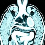
CT scanning is often the first-line imaging modality for many acute abdominal pain presentations, particularly when a quick and comprehensive assessment is needed. It uses X-rays to create detailed cross-sectional images of the abdomen and pelvis. The speed with which CT scans can be performed makes them invaluable in emergency situations where rapid diagnosis is crucial, such as suspected appendicitis, bowel obstruction, or internal bleeding.
– High resolution allows for visualization of both bony structures and soft tissues.
– Relatively fast scan times are ideal for patients who are unable to remain still for extended periods.
However, CT scans do involve exposure to ionizing radiation, which is a concern, especially in children and pregnant women. Contrast agents (often iodine-based) are frequently used to enhance image clarity, but these can pose risks to individuals with kidney problems or allergies. Modern protocols increasingly focus on minimizing contrast use and radiation dose while maintaining diagnostic quality.
CT scans excel at identifying conditions like:
* Appendicitis: Inflammation of the appendix.
* Diverticulitis: Infection/inflammation of small pouches in the colon.
* Kidney stones: Calcifications within the urinary tract.
* Bowel obstruction: Blockage preventing normal passage of intestinal contents.
* Mesenteric ischemia: Reduced blood flow to the intestines.
* Trauma-related injuries: Detecting internal bleeding or organ damage.
For children experiencing abdominal discomfort, it’s important to understand when belly pain is your child’s way of saying help.
Magnetic Resonance Imaging (MRI) Scans
MRI utilizes strong magnetic fields and radio waves to generate detailed images, offering excellent soft tissue contrast without ionizing radiation. This makes it a valuable alternative to CT scanning in certain situations, particularly for patients who require repeated imaging or are concerned about radiation exposure. While MRI provides superior visualization of soft tissues – including the liver, pancreas, gallbladder, bile ducts, and bowel wall– it generally takes longer than a CT scan and can be less readily available.
– No ionizing radiation is a significant advantage.
– Excellent soft tissue detail allows for better assessment of certain conditions.
However, MRI scans are more expensive than CT scans, may be contraindicated in patients with metallic implants (pacemakers, some aneurysm clips), and can be challenging for claustrophobic individuals.
MRI is particularly useful for:
* Evaluating liver disease and tumors.
* Assessing the pancreas and bile ducts.
* Diagnosing Crohn’s disease and other inflammatory bowel conditions.
* Characterizing complex abdominal masses.
* Identifying subtle injuries not visible on CT scans.
Ultrasound Examination
Advantages and Limitations
Ultrasound uses sound waves to create images of internal organs, offering a non-invasive, relatively inexpensive, and readily available diagnostic tool. It’s particularly useful for evaluating the gallbladder, liver, kidneys, and pancreas, as well as assessing blood flow in major vessels. A key advantage is its lack of ionizing radiation, making it safe for pregnant women and children. However, ultrasound image quality can be affected by factors like body habitus (patient size), bowel gas, and operator skill. It also doesn’t penetrate bone well, limiting its ability to visualize structures behind bony tissues.
– Real-time imaging allows for dynamic assessment of organ function.
– Portability makes it convenient for bedside examinations.
– Cost-effective compared to CT and MRI scans.
Common Applications in Abdominal Pain
Ultrasound is frequently used as an initial screening tool for:
* Gallstones: Stones forming within the gallbladder.
* Cholecystitis: Inflammation of the gallbladder, often caused by gallstones.
* Kidney stones (smaller stones).
* Liver abnormalities: Detecting cysts, tumors, or fluid collections.
* Evaluating appendicitis in children (although CT is still preferred for adults)
* Assessing blood flow to organs.
Managing discomfort after a meal? Learn about post-meal rituals that help prevent abdominal bloating.
Endoscopic Ultrasound (EUS)
EUS combines the principles of endoscopy and ultrasound, allowing for detailed imaging of the gastrointestinal tract and surrounding structures from within the body. A flexible endoscope with an attached ultrasound probe is inserted through the mouth or rectum, providing high-resolution images of the esophagus, stomach, duodenum, pancreas, gallbladder, bile ducts, and rectum.
– High resolution provides detailed visualization of pancreatic and biliary structures.
– Biopsy capability allows for tissue sampling during the procedure.
EUS is particularly valuable in diagnosing:
1. Pancreatic cancer and cysts.
2. Biliary tract disorders.
3. Gastrointestinal stromal tumors (GIST).
4. Inflammatory bowel disease.
Nuclear Medicine Scans
Nuclear medicine scans involve injecting a small amount of radioactive tracer into the body, which is then detected by a special camera to create images that reflect organ function. These scans are less about anatomical detail and more about assessing physiological processes. For example, a hepatobiliary scan (HIDA scan) can evaluate gallbladder function and detect blockages in the bile ducts.
– Functional imaging provides information about how organs are working.
– Can detect subtle abnormalities not visible on other scans.
Nuclear medicine scans are useful for:
1. Detecting infections or inflammation.
2. Assessing liver function.
3. Identifying gastrointestinal bleeding sources.
4. Evaluating the biliary system (HIDA scan).
For relief from gas and discomfort, consider abdominal stretch routines for releasing trapped gas. If you are experiencing significant pain or have concerns, it is critical to know when to seek medical attention for abdominal pain. Understanding your digestive habits can also play a role in managing discomfort – explore digestive habits that help during high travel periods. Finally, remember to consider the power of food rituals that help your gut feel safe and steady
The choice of which scan to use ultimately depends on a careful assessment of the patient’s individual situation and the suspected cause of their abdominal pain. A skilled healthcare professional will consider all available information, including medical history, physical examination findings, laboratory tests, and imaging results, to arrive at an accurate diagnosis and develop an appropriate treatment plan. It is vital to discuss any concerns or questions you have about recommended scans with your physician.


