Upper endoscopy, also known as esophagogastroduodenoscopy (EGD), is a remarkably common procedure used to visually examine the upper digestive tract. It’s often recommended when someone experiences persistent symptoms like heartburn, difficulty swallowing, abdominal pain, nausea, vomiting, or unexplained weight loss. But what exactly do doctors see during this process? Beyond simply looking for obvious problems, an endoscopist is trained to recognize subtle signs of disease and identify areas requiring further investigation. The procedure itself involves a flexible tube with a camera attached being gently passed down the esophagus, into the stomach, and finally into the duodenum – the first part of the small intestine. This article will explore what doctors are looking for during an upper endoscopy, offering insight into the anatomy observed and common findings encountered.
The goal isn’t just to diagnose; it’s to gain a comprehensive understanding of the patient’s digestive health. An endoscopist is essentially performing a real-time visual inspection, assessing not only the structure of the organs but also the surface characteristics, looking for abnormalities in color, texture, and shape. This allows them to differentiate between benign conditions and those that might require intervention or further testing such as biopsies. Understanding what doctors observe can alleviate anxiety for patients undergoing the procedure and provide a better appreciation of its diagnostic power. It’s important to remember that endoscopy is often preventative – identifying problems early before they become more serious complications. If you experience reflux only during travel, understanding these procedures might ease concerns.
Anatomy Observed During Upper Endoscopy
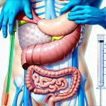
The upper digestive tract consists of several key structures, each with distinct characteristics an endoscopist evaluates. The esophagus is the first stop, a muscular tube connecting the throat to the stomach. Its lining should be smooth and pink, without any signs of inflammation or ulceration. As the scope enters the stomach, the environment changes significantly. The stomach’s mucosal lining appears as a series of folds called rugae, which allow it to expand after eating. Doctors scrutinize these rugae for irregularities like redness, swelling, or erosions. Finally, the duodenum – the shortest part of the small intestine – is examined. Here, the focus shifts to the ampulla of Vater, where bile from the liver and pancreatic enzymes enter the digestive process, and the duodenal bulb which is often prone to ulceration.
The endoscopist isn’t just looking at these organs, they are assessing their relationships to each other and identifying any structural anomalies. For example, a hiatal hernia – where part of the stomach protrudes through the diaphragm – would be readily visible during esophageal examination. Similarly, the presence of varices (enlarged veins) in the esophagus or stomach can indicate portal hypertension, often linked to liver disease. The entire journey is performed under direct visualization on a monitor, allowing for meticulous assessment and documentation of findings. Understanding what happens to your esophagus during chronic heartburn is key to preventative care.
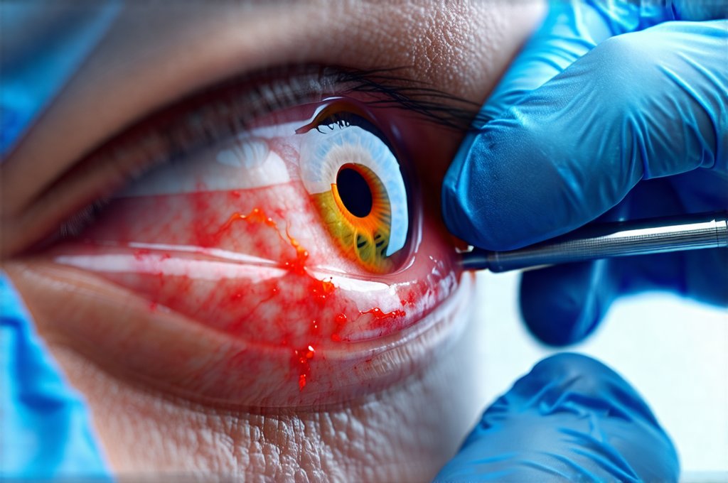
The color changes observed are crucial diagnostic clues. Pale areas might suggest bleeding or anemia, while bright red indicates active inflammation or ulcers. A yellowish tinge could signal bile reflux. Furthermore, the texture of the lining provides vital information – smooth is generally healthy, but rough or nodular textures raise suspicion for pre-cancerous conditions or cancerous growths. The ability to clearly differentiate between normal and abnormal tissue is what makes upper endoscopy such a valuable diagnostic tool.
Common Findings & What They Indicate
During an upper endoscopy, doctors frequently encounter several common findings which range in severity. Gastritis, inflammation of the stomach lining, is one of the most prevalent discoveries. It often appears as redness and swelling and can be caused by factors like H. pylori infection, NSAID use, or excessive alcohol consumption. Another frequent finding is esophagitis, an inflammation of the esophagus, commonly due to acid reflux (GERD). This typically manifests as redness, ulceration, or even narrowing of the esophageal lumen.
Peptic ulcers – sores in the lining of the stomach or duodenum – are also relatively common. They appear as distinct craters and can cause significant pain and bleeding. Doctors will assess their size, location, and depth to determine appropriate treatment. Importantly, endoscopy allows for biopsies to be taken from these ulcers to rule out malignancy (cancer). It’s important to note that many individuals may have polyps – small growths in the stomach or colon – which are generally benign but require evaluation through biopsy to exclude cancerous changes. Knowing what really triggers heartburn even on an empty stomach can help you prepare for a visit.
Beyond these common findings, endoscopists also look for more subtle signs of disease. These include: – Subtle changes in mucosal pattern – Small erosions or ulcers that might not cause obvious symptoms – The presence of bleeding points – Evidence of previous surgeries (scars) – Foreign bodies (rarely). The ability to identify these subtle clues often leads to earlier diagnosis and intervention, improving patient outcomes. Considering an anti-inflammatory diet may help prevent some findings.
Biopsies & Further Investigation
A critical component of upper endoscopy is the ability to take biopsies. A biopsy involves removing a small sample of tissue for microscopic examination by a pathologist. This is essential for diagnosing conditions like H. pylori infection (a common cause of gastritis and ulcers), Barrett’s esophagus (a pre-cancerous condition in the esophagus caused by chronic acid reflux), and various types of cancers. Biopsies are typically painless, as the lining of the digestive tract doesn’t have many nerve endings.
The doctor will use instruments passed through the endoscope to obtain biopsies from any suspicious areas or lesions identified during the examination. Multiple biopsies may be taken from different locations within a lesion to ensure accurate diagnosis. After the biopsies are collected, they’re sent to a laboratory for analysis and results typically take several days to become available.
If endoscopy reveals significant abnormalities like large polyps or suspected cancer, further investigation might be necessary. This could include: 1) CT scans or MRI to assess the extent of disease 2) Additional endoscopic procedures such as ERCP (endoscopic retrograde cholangiopancreatography) to examine the bile ducts and pancreatic duct 3) Surgical intervention for removal of larger tumors or lesions. The decision regarding further investigation is always tailored to the individual patient and their specific findings. If you experience GERD and sudden chills during an episode, it’s best to consult a doctor.
The ultimate goal of upper endoscopy isn’t just identifying problems, but providing a clear path forward in terms of treatment and management. By combining visual assessment with targeted biopsies, doctors can provide accurate diagnoses and develop personalized care plans that optimize patient outcomes. When considering dietary changes, knowing what to avoid on an anti-inflammatory diet is helpful.

