Abdominal pain, bloating, nausea, changes in bowel habits – these are all incredibly common complaints that physicians encounter daily. The sheer breadth of potential causes, ranging from minor indigestion to serious underlying disease, makes initial assessment a significant challenge. Patients often present with vague symptoms, making diagnosis difficult without a systematic approach. A crucial first step in investigating these abdominal issues is often imaging, and increasingly, ultrasound is becoming the modality of choice for many clinicians due to its accessibility, relatively low cost, non-invasive nature and lack of ionizing radiation. This article will explore the role of ultrasound as that initial investigative tool, detailing what it can reveal, its limitations, and how it fits into a broader diagnostic pathway.
The use of ultrasound allows healthcare professionals to quickly gather valuable information about the abdominal organs without subjecting patients to more complex or potentially harmful investigations like CT scans or MRI. It’s particularly useful in triaging patients – helping determine whether urgent intervention is needed or if further observation and less invasive tests are appropriate. While not always definitive, an initial ultrasound can significantly narrow down the differential diagnosis, leading to faster and more accurate treatment plans. Importantly, it’s a dynamic examination; meaning the sonographer can assess organs both statically and in real-time, observing how they function and respond to pressure or manipulation during the scan. Considering other conditions with similar symptoms, like gerd vs ulcer, can also aid in diagnosis.
The Power of Ultrasound Imaging
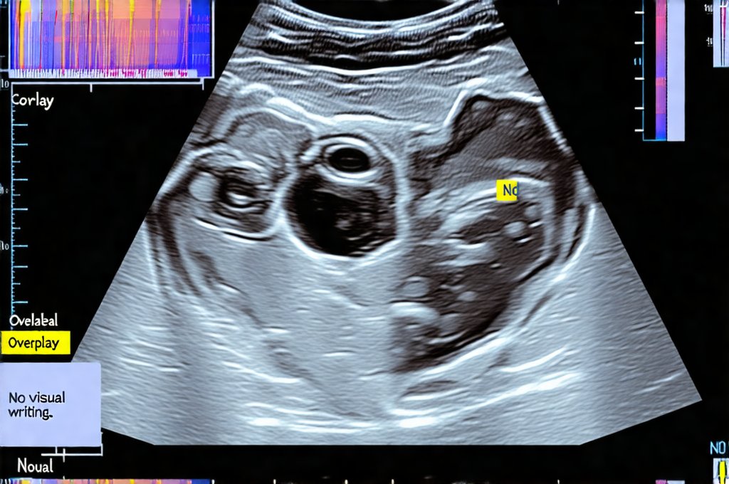
Ultrasound utilizes high-frequency sound waves to create images of the structures within the abdomen. A transducer emits these sound waves, which penetrate the body and are reflected back differently depending on the density of the tissues they encounter. These reflections are then processed into a visual image displayed on a screen. Different types of ultrasound techniques exist, but for abdominal imaging, several key features make it particularly useful: – It’s readily available in most hospitals and clinics. – It doesn’t require contrast agents (although some specialized scans may use them). – It provides real-time imaging, allowing assessment of blood flow and organ movement. – This is often referred to as Doppler ultrasound.
The ability to visualize key abdominal organs makes ultrasound invaluable in identifying a wide range of conditions. For instance, gallbladder disease (such as gallstones), liver abnormalities, kidney stones, and even some bowel obstructions can be detected with reasonable accuracy using this technique. It’s also extremely useful in guiding procedures like biopsies or fluid aspirations, ensuring precision and minimizing complications. However, it’s important to remember that ultrasound is operator-dependent; the skill and experience of the sonographer directly impact the quality of the images obtained. Understanding breathing techniques can also help manage related discomforts during examination.
Ultrasound isn’t without limitations. Air and bone can obstruct sound waves, making visualization of certain areas – particularly behind the bowel or in obese patients – challenging. It also doesn’t provide detailed imaging of structures deep within the abdomen as effectively as CT or MRI. Therefore, it often serves as a ‘first line’ investigation, guiding subsequent more advanced imaging if necessary. The quality of images can vary significantly based on patient body habitus and the presence of bowel gas. This means that while highly useful, ultrasound findings should always be interpreted in conjunction with clinical assessment and other relevant investigations. If symptoms are worse in the morning, it can influence diagnostic approach.
Ultrasound for Specific Abdominal Complaints
When a patient presents with abdominal pain, the initial focus is often to rule out life-threatening conditions. Ultrasound can quickly assess for: – Appendicitis: While CT remains the gold standard, ultrasound can sometimes identify signs of appendiceal inflammation, especially in children and pregnant women. – Cholecystitis (Gallbladder Inflammation): This is a very common indication for abdominal ultrasound. The presence of gallstones or thickening of the gallbladder wall can strongly suggest cholecystitis. – Often accompanied by tenderness to palpation over the right upper quadrant. – Pancreatitis: Ultrasound can detect pancreatic inflammation and identify fluid collections around the pancreas, although CT is often preferred for a more detailed assessment. It’s important to consider conditions like bile reflux that can mimic similar symptoms.
The use of Doppler ultrasound significantly enhances diagnostic capabilities. By measuring blood flow, it can help determine if a blockage exists in an artery or vein. For example, in suspected cases of mesenteric ischemia (reduced blood flow to the intestines), Doppler ultrasound can assess arterial and venous patency. Similarly, in patients with abdominal aortic aneurysm, Doppler can be used to monitor the size and stability of the aneurysm. The dynamic nature of ultrasound allows for a more comprehensive evaluation than static imaging techniques. Understanding histamine’s role in abdominal discomfort can also be beneficial.
Furthermore, ultrasound is exceptionally useful in assessing the liver. It can detect fatty liver disease, cirrhosis, tumors, and abscesses. While MRI or CT are usually required for detailed characterization of liver lesions, ultrasound provides an initial screening tool to identify abnormalities that warrant further investigation. In cases of jaundice (yellowing of the skin), ultrasound can help determine if the obstruction is within the bile ducts, potentially indicating a gallstone or tumor. It’s often used as part of the workup for unexplained weight loss and changes in liver function tests.
The Role of Ultrasound in Bowel Investigation
While ultrasound isn’t ideal for visualizing the entire length of the bowel due to gas interference, it can be useful in specific scenarios. Identifying small bowel obstructions is one example. Although CT scans are generally preferred, ultrasound can often detect dilated loops of bowel and identify potential causes, like adhesions or hernias. In patients with suspected inflammatory bowel disease (IBD), such as Crohn’s disease or ulcerative colitis, ultrasound can assess the thickness of the intestinal wall and detect areas of inflammation. – This is often referred to as intestinal wall thickening. Dietary considerations, like gluten’s impact, may also be explored concurrently.
In children, ultrasound is frequently used to diagnose intussusception, a condition where one part of the intestine slides into another. It’s a relatively safe and accurate method for identifying this condition, which can cause significant abdominal pain and vomiting in infants. Ultrasound can also help differentiate between various causes of diarrhea and abdominal cramping, although stool studies are often necessary to confirm the diagnosis. The ability to visualize fluid collections within the abdomen is also valuable in detecting complications related to bowel perforation or abscess formation. Peppermint oil can sometimes offer relief from associated symptoms.
Finally, ultrasound plays a crucial role in guiding percutaneous drainages for intra-abdominal abscesses. By providing real-time visualization, it ensures that the drainage catheter is placed accurately and safely, minimizing the risk of damage to surrounding organs. This is often performed under sterile conditions with appropriate imaging guidance and antibiotic coverage. It’s an excellent example of how ultrasound isn’t just a diagnostic tool but can also be integral to therapeutic interventions.
It is essential to remember that this information is for general knowledge and informational purposes only, and does not constitute medical advice. Always consult with a qualified healthcare professional for any health concerns or before making any decisions related to your health or treatment.

