Hidden bleeding, often termed “occult” or internal bleeding, presents a significant diagnostic challenge in clinical medicine. Unlike overt bleeding – such as from a visible wound – occult bleeding may not have immediately obvious symptoms, making its detection reliant on astute observation and increasingly, advanced medical imaging techniques. This can range from slow, chronic blood loss leading to anemia to acute, life-threatening hemorrhages occurring within the body. Early and accurate identification of these bleeds is paramount for effective treatment and improved patient outcomes; delays in diagnosis can lead to significant morbidity and mortality. The complexity lies not only in identifying where the bleeding originates but also in determining its rate and source, often requiring a multifaceted approach utilizing various imaging modalities.
The challenge is further compounded by the fact that symptoms of occult bleeding are frequently non-specific, mimicking other conditions. Fatigue, weakness, shortness of breath, dizziness, and paleness can all be indicative, but are not exclusive to internal hemorrhage. The patient may have no immediately apparent external signs, making diagnosis dependent on clinical suspicion based on risk factors (e.g., anticoagulant use, gastrointestinal history) or incidental findings during routine testing (e.g., anemia detected in a blood test). This necessitates a robust arsenal of imaging tools capable of visualizing internal structures with high sensitivity and specificity to pinpoint the source of bleeding when other methods are insufficient. Considering potential hidden triggers can also aid diagnosis.
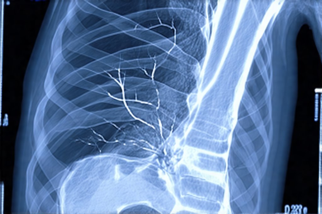
The Role of Computed Tomography (CT) Scanning
Computed tomography, commonly known as CT scanning, is a mainstay in emergency medicine and plays a critical role in identifying acute intra-abdominal and intrathoracic bleeding. Utilizing X-rays, CT scans create detailed cross-sectional images of the body, allowing clinicians to visualize organs, blood vessels, and any associated abnormalities. CT’s speed and accessibility make it invaluable when rapid diagnosis is crucial. While traditional CT can detect significant hemorrhage, advancements in technology have enhanced its capabilities for detecting subtle bleeding.
Contrast-enhanced CT (CECT) significantly improves visualization of vascular structures. Intravenous contrast agents containing iodine highlight blood vessels, making it easier to identify areas of extravasation—where blood leaks out from a vessel. However, CECT isn’t without limitations; patients with kidney disease or allergies to iodine may not be suitable candidates. Newer techniques like dual-energy CT (DECT) are gaining traction as they can differentiate between different tissues based on their energy absorption properties, potentially improving the detection of even small amounts of bleeding without relying solely on contrast agents.
Furthermore, CT angiography (CTA), a specialized form of CECT focused specifically on blood vessels, is particularly useful for identifying active bleeding sites within arteries and veins. A CTA can rapidly assess for vascular injury following trauma or identify aneurysms that may be leaking. The interpretation of CT scans requires skilled radiologists who can differentiate between normal anatomy, acute bleeding, old blood products (which appear different), and other potential causes of symptoms. It’s vital to also consider hidden ingredients in food as a contributing factor to overall health.
Ultrasound’s Complementary Role in Bleeding Detection
While CT is powerful for broad anatomical assessment, ultrasound offers a non-invasive and readily available alternative, especially in certain clinical scenarios. It’s particularly useful in emergency settings for rapid initial assessment due to its portability and lack of ionizing radiation. Focused Assessment with Sonography for Trauma (FAST) exam utilizes ultrasound to quickly identify free fluid in the abdomen – often indicating internal bleeding after trauma.
FAST exams are typically performed in patients presenting with abdominal injuries following blunt or penetrating trauma.
The technique involves scanning specific anatomical locations, including around the liver, spleen, and pelvis, looking for signs of fluid accumulation (hypoechoic areas).
Ultrasound is also used to assess for pericardial effusion, which can indicate bleeding around the heart.
However, ultrasound has limitations. Its ability to penetrate deeper tissues can be limited by body habitus or bowel gas, making it less effective in obese patients or those with significant intestinal distension. It’s also operator dependent – accurate interpretation requires skilled sonographers and clinicians trained in ultrasound imaging. Despite these limitations, the speed and accessibility of ultrasound make it an invaluable tool for initial triage and assessment, often guiding subsequent investigations such as CT scans. Understanding hidden GERD symptoms can help with differential diagnosis.
Magnetic Resonance Imaging (MRI) for Complex Cases
Magnetic resonance imaging (MRI) provides exceptional soft tissue detail and doesn’t involve ionizing radiation, making it a valuable adjunct in diagnosing certain types of occult bleeding, particularly when other modalities are inconclusive or have limitations. While not typically the first-line imaging modality in acute settings due to its longer scan times, MRI excels at identifying chronic bleeding, such as small hemorrhages within the brain or spinal cord.
MRI offers several advantages over CT and ultrasound in specific situations:
– It can differentiate between acute and subacute/chronic blood products with greater accuracy. This is crucial for determining the timing of a bleed and guiding treatment decisions.
– Gradient echo MRI sequences are highly sensitive to detecting even microscopic bleeding, making them useful in evaluating patients with suspected cerebral microbleeds or spinal cord injuries.
– Susceptibility Weighted Imaging (SWI) is an advanced MRI technique that enhances visualization of blood products, further improving detection rates.
However, MRI is more expensive than CT and ultrasound, and it’s contraindicated in patients with certain metallic implants. The longer scan times can also be a disadvantage in unstable patients requiring immediate intervention. Therefore, the decision to utilize MRI for bleeding detection should be based on careful consideration of the clinical context, patient factors, and available resources. Additionally, watch hidden ingredients to watch when considering dietary impacts.
It is important to note that advancements continue in all areas of medical imaging, leading to improved sensitivity, specificity, and reduced radiation exposure. These technologies are constantly evolving, providing clinicians with increasingly sophisticated tools to detect and manage occult bleeding effectively. Ultimately, a collaborative approach involving radiologists, emergency physicians, and other specialists is essential for accurate diagnosis and optimal patient care. Don’t overlook the hidden role of dehydration in exacerbating these conditions. Also, consider if there are hidden triggers in supplements that could be contributing.
Dr. Olivia Bennett, MD is a board-certified gastroenterologist specializing in advanced digestive diagnostics. With over 12 years of clinical experience, she focuses on non-invasive testing methods, gut microbiome assessment, and functional GI disorders. Dr. Bennett is passionate about helping patients understand their digestive health through clear and accessible medical insights.