Initial scans used before more invasive digestive tests
Digestive health is often something we take for granted until an issue arises. Discomfort, changes in bowel habits, persistent heartburn – these can all signal underlying problems within our complex digestive system. Diagnosing those problems effectively requires a careful and methodical approach, rarely beginning with the most invasive tests. Instead, healthcare professionals typically start with less intrusive initial scans and assessments to narrow down potential causes before proceeding to more detailed investigations like colonoscopies or endoscopies. This staged approach minimizes patient discomfort, reduces costs, and ensures that only those who truly require further investigation undergo potentially stressful procedures. The goal isn’t just finding a problem, but understanding it accurately and efficiently.
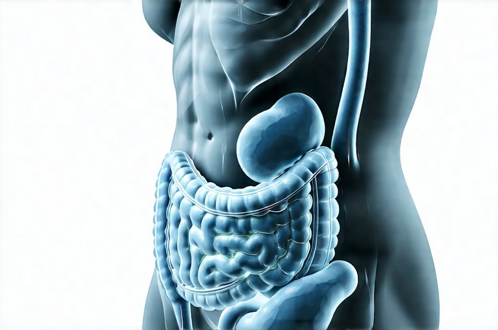
The initial evaluation process is crucial because the digestive system encompasses a wide range of organs – esophagus, stomach, intestines (both small and large), liver, gallbladder, pancreas – each with its own potential for dysfunction. Symptoms can often be vague or overlap between different conditions. For example, abdominal pain could stem from irritable bowel syndrome, gallstones, pancreatitis, or even a heart condition! Therefore, initial scans act as a ‘first filter’, helping doctors differentiate between possibilities and direct resources toward the most likely diagnosis. A well-chosen sequence of preliminary tests can prevent unnecessary procedures and guide treatment strategies effectively. Understanding your food energy patterns is also an important first step.
Non-Invasive Imaging Techniques
Initial imaging techniques play a pivotal role in assessing digestive health without requiring incisions or internal instrumentation. These methods offer valuable insights into the structure and function of various organs, providing clues about potential abnormalities. X-rays are often among the first lines of investigation due to their accessibility and relatively low cost. However, they have limitations regarding soft tissue visualization. More advanced imaging modalities like CT scans and MRIs provide considerably more detail and are employed when greater precision is needed. Ultrasound is another valuable tool, particularly for evaluating gallbladder issues or detecting fluid accumulation. If you struggle with knowing how to eat more without discomfort, initial scans can help pinpoint the cause.
CT (Computed Tomography) scans utilize X-rays but create cross-sectional images of the body, offering a much clearer view than traditional X-rays. They’re excellent for identifying tumors, abscesses, inflammation, and blockages within the digestive tract. A CT scan with contrast – where a dye is injected intravenously – further enhances visualization by highlighting blood vessels and organs. MRI (Magnetic Resonance Imaging) uses strong magnetic fields and radio waves to produce detailed images, excelling at soft tissue imaging, making it particularly useful for evaluating liver, pancreas, and bowel disorders. While MRIs avoid radiation exposure, they tend to be more expensive and time-consuming than CT scans.
Ultrasound utilizes sound waves to create real-time images of organs. It’s frequently used to assess the gallbladder for gallstones, evaluate the liver for abnormalities, and detect fluid in the abdomen. It’s non-invasive, relatively inexpensive, and doesn’t involve radiation, making it a safe option for many patients. However, its image quality can be affected by factors like bowel gas or patient body habitus. The choice of imaging modality depends on the specific symptoms, suspected diagnosis, and individual patient characteristics. Creating daily eating maps can help you understand how your body reacts to different foods.
Blood Tests & Stool Analysis
Beyond imaging, laboratory tests provide essential information about digestive health. Blood tests are fundamental, often starting with a complete blood count (CBC) to assess overall health and look for signs of infection or inflammation. Liver function tests (LFTs) evaluate liver health, while pancreatic enzyme levels can indicate pancreatitis. Specific biomarkers can also help identify conditions like celiac disease or inflammatory bowel disease (IBD). A seemingly simple blood test can be incredibly informative in guiding the diagnostic process.
Stool analysis is another valuable non-invasive tool. It can detect the presence of blood (indicating bleeding), parasites, bacteria, or abnormal levels of fat (suggesting malabsorption). Fecal immunochemical tests (FIT) are specifically designed to screen for colorectal cancer by detecting microscopic amounts of blood in stool. More advanced stool tests can also identify inflammatory markers, providing clues about IBD. These tests are relatively easy to perform at home and offer a convenient way to gather important diagnostic information. For those looking to build comfort-based food routines, stool analysis can help identify trigger foods.
Endoscopic Capsule
The endoscopic capsule represents a significant advancement in non-invasive digestive diagnostics. This tiny wireless camera is swallowed by the patient and travels through the entire small intestine, taking thousands of images along the way. These images are then transmitted wirelessly to a recorder worn by the patient. This technology allows for visualization of areas that are difficult or impossible to reach with traditional endoscopy – such as the small intestine which can be challenging to access.
The capsule is painless and generally well-tolerated, although it requires some dietary preparation before ingestion.
It’s particularly useful in identifying sources of bleeding, detecting tumors, and evaluating inflammatory conditions like Crohn’s disease.
While incredibly valuable, the capsule doesn’t allow for biopsies or therapeutic interventions; if abnormalities are found, further investigation with traditional endoscopy is often required. The capsule provides a comprehensive visual assessment without the discomfort associated with more invasive procedures, making it a powerful diagnostic tool. Understanding meal timelines can help you prepare for this type of scan.
It’s important to remember that these initial scans are rarely definitive on their own. They provide crucial starting points for diagnosis but often need to be combined with other tests and clinical evaluation to arrive at an accurate conclusion. The process is collaborative – doctors carefully interpret the results of each test, considering the patient’s symptoms, medical history, and physical examination findings to develop a personalized diagnostic plan. Many find that prep-ahead meals simplify dietary preparation for scans and tests.
Dr. Olivia Bennett, MD is a board-certified gastroenterologist specializing in advanced digestive diagnostics. With over 12 years of clinical experience, she focuses on non-invasive testing methods, gut microbiome assessment, and functional GI disorders. Dr. Bennett is passionate about helping patients understand their digestive health through clear and accessible medical insights.