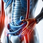
Our digestive system is arguably one of the most crucial systems in our bodies, responsible for breaking down food into nutrients our cells can use for energy, growth, and repair. When this intricate process goes awry, it can manifest as a wide range of uncomfortable and sometimes debilitating symptoms impacting daily life. Many people experience occasional digestive discomfort – bloating after a large meal or temporary constipation – but persistent or severe issues often signal underlying problems that require investigation. Fortunately, advancements in medical imaging technology now allow for relatively non-invasive assessment of many common digestive concerns, offering quicker diagnoses and more targeted treatments than ever before.
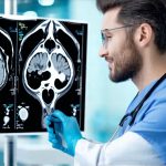
Historically, diagnosing digestive ailments relied heavily on subjective patient reporting, invasive procedures like endoscopy and colonoscopy, or lengthy waiting periods for symptom patterns to emerge. Today, a variety of scanning techniques – from readily available ultrasound to sophisticated CT and MRI scans – can provide visual information about the structures and functions within our digestive tract. This isn’t about replacing traditional diagnostic methods; rather, it’s about adding another powerful tool to the physician’s arsenal, improving accuracy, reducing patient discomfort, and accelerating the path toward effective care. We will explore some of the key digestive problems that can be detected with these relatively simple scans, highlighting what each scan reveals and how it aids in diagnosis.
Imaging for Structural Abnormalities
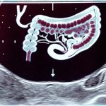
Many digestive issues stem from physical changes within the digestive organs themselves – growths, blockages, inflammation, or structural defects. Scanning technologies excel at identifying these abnormalities. Ultrasound is often a first-line investigation due to its affordability, accessibility, and lack of ionizing radiation. It’s particularly useful for examining the gallbladder, detecting gallstones which are a common cause of abdominal pain, and assessing the liver for signs of disease. Computed Tomography (CT) scans offer more detailed cross-sectional images, allowing doctors to visualize the entire digestive tract – from esophagus to rectum – and identify problems like tumors, diverticulitis (inflammation or infection in small pouches that can form in the lining of the colon), appendicitis, and bowel obstructions. Magnetic Resonance Imaging (MRI), while generally more expensive and time-consuming than CT scans, provides even greater soft tissue detail, making it ideal for evaluating inflammatory bowel disease (IBD) like Crohn’s disease and ulcerative colitis, as well as detecting subtle tumors or lesions that might be missed on a CT scan.
The benefit of these imaging methods lies not just in identifying the problem but also in ruling out others. For instance, abdominal pain could have numerous causes – gynecological issues, kidney stones, musculoskeletal problems – and an initial scan can help narrow down the possibilities, directing further investigation more efficiently. Moreover, scans allow for assessment of the extent of a disease; is a tumor localized or has it spread? How severe is the inflammation in the bowel? This information is crucial for treatment planning. It’s also helpful to understand how [digestive exams that can be done without sedation] may fit into your overall diagnostic plan.
Crucially, these scans don’t always provide definitive diagnoses on their own. Often, they highlight areas of concern that require further investigation through endoscopy or biopsy. However, they significantly streamline the diagnostic process and reduce the need for exploratory surgery. The increasing availability of 3D reconstruction from CT and MRI data also allows surgeons to plan operations with greater precision, improving outcomes and minimizing risks. If you are facing surgery, it is important to consider [digestive tests that should be done before surgery].
Detecting Gallbladder Issues
The gallbladder is a small organ that stores bile produced by the liver. Problems within the gallbladder are incredibly common, often presenting as sudden, intense pain in the upper right abdomen. Ultrasound is the go-to imaging modality for evaluating gallbladder issues. – It can readily identify gallstones, which are hardened deposits of digestive fluid; these stones can block the cystic duct (the tube connecting the gallbladder to the bile duct), causing inflammation and excruciating pain known as a biliary colic. – Furthermore, ultrasound can detect signs of cholecystitis, or inflammation of the gallbladder itself, often caused by prolonged blockage from gallstones. Signs include thickening of the gallbladder wall and fluid around the gallbladder.
Beyond simple gallstones, scans can also reveal polyps within the gallbladder – small growths that are usually benign but occasionally warrant further investigation to rule out malignancy. More sophisticated imaging like MRCP (magnetic resonance cholangiopancreatography) provides detailed views of the bile ducts, helping identify blockages or narrowing caused by stones, tumors, or inflammation. This allows for precise planning of treatment, ranging from medication to dissolve smaller stones to surgical removal of the gallbladder (cholecystectomy). Understanding [food energy patterns that align with digestive clarity] can also help manage your symptoms.
Identifying Bowel Obstructions and Inflammation
Bowel obstructions can occur due to a variety of reasons – adhesions from previous surgery, hernias, tumors, or inflammatory bowel disease. CT scans are exceptionally effective in identifying these blockages. – They show where the blockage is located, what’s causing it (e.g., a tumor pressing on the bowel), and whether there’s any associated fluid buildup or bowel wall thickening. This information is vital for determining the urgency of treatment; complete obstructions require immediate intervention to prevent serious complications like bowel perforation.
Inflammation within the bowel, as seen in conditions like Crohn’s disease and ulcerative colitis, can also be detected on CT scans and MRI. – While endoscopy remains the gold standard for visualizing the inner lining of the bowel, imaging provides a broader overview of inflammation extending deeper into the bowel wall. MRI, in particular, excels at differentiating between active inflammation and chronic scarring, helping guide treatment decisions. A key feature visible on scan is bowel wall thickening, indicating an inflammatory response. [Digestive investigations that don’t require a hospital visit] are also available for less severe cases.
Evaluating Appendicitis and Diverticulitis
Appendicitis, or inflammation of the appendix, often presents with right lower quadrant abdominal pain. While clinical evaluation remains important, CT scans are now routinely used to confirm the diagnosis – they can show a swollen appendix, fluid around it, and sometimes even the presence of an appendicolith (a hardened fecal mass within the appendix). This helps avoid unnecessary surgery in cases where the symptoms might be due to other causes. Diverticulitis, as previously mentioned, involves inflammation or infection of small pouches that form in the colon. – CT scans are essential for diagnosing diverticulitis because they can show thickened bowel wall, inflammation around the diverticula (the pouches), and potential complications like abscesses or perforations.
It’s important to note that not all patients with diverticula have diverticulitis; many people have these pouches without experiencing any symptoms. Scans help differentiate between asymptomatic diverticulosis and symptomatic diverticulitis requiring treatment, typically antibiotics and dietary changes. [Home kits that can detect digestive infections] may also be useful in some situations. If you are concerned about underlying issues, it’s important to determine [can blood tests detect digestive problems].
The information provided in this article is for general knowledge and informational purposes only, and does not constitute medical advice. It is essential to consult with a qualified healthcare professional for any health concerns or before making any decisions related to your health or treatment.


