Common scans doctors use to explore digestive pain
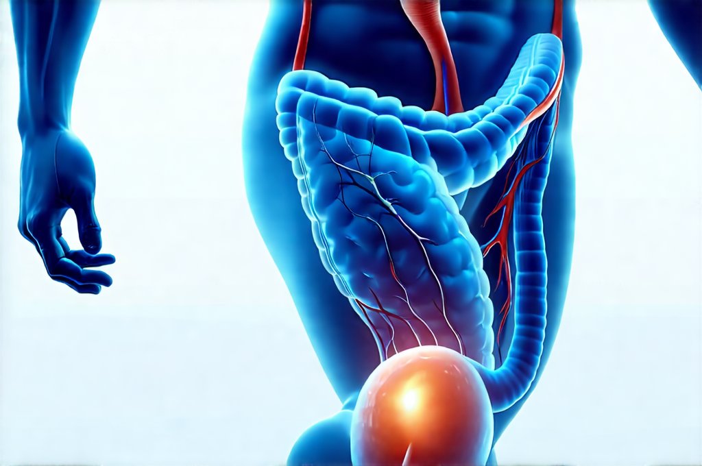
Digestive pain is an incredibly common complaint, impacting millions worldwide. It’s rarely a single issue; rather, it’s often a symptom stemming from a vast range of potential causes – from relatively minor issues like gas and bloating to more serious conditions affecting the esophagus, stomach, intestines, liver, gallbladder, or pancreas. Because of this complexity, diagnosing the source of digestive pain requires careful investigation, and imaging plays a crucial role in that process. Doctors employ a diverse toolkit of scans and tests to visualize the internal workings of the digestive system, pinpointing problem areas with increasing accuracy. Understanding what these scans are, how they work, and when doctors choose them can empower patients to better understand their own care.
The selection of which scan to use depends heavily on the specific symptoms a patient is experiencing, as well as initial physical examinations and medical history. A doctor will consider factors like where the pain is located (upper abdomen, lower abdomen, etc.), its character (sharp, dull, cramping), what makes it better or worse, and any accompanying symptoms such as nausea, vomiting, diarrhea, constipation, or weight loss. It’s important to remember that scans are usually part of a larger diagnostic process – they rarely provide the complete answer on their own. They often guide further testing, like endoscopies or biopsies, to confirm a diagnosis. This article will explore some of the most common scanning methods used to investigate digestive pain, providing insight into what patients can expect during these procedures and how the results are interpreted.
Imaging the Digestive Tract: X-rays and CT Scans
X-rays have been a cornerstone of medical imaging for over a century, and remain valuable in initial assessments of digestive issues. While they don’t offer the detailed views of more modern techniques, they’re quick, inexpensive, and readily available. A typical abdominal X-ray can help identify blockages, perforations (holes), or large masses in the digestive tract. However, soft tissues – like the intestines themselves – are difficult to see clearly on a standard X-ray. To improve visualization, doctors often use barium contrast studies. This involves patients drinking a barium solution which coats the lining of the esophagus, stomach, and small intestine, making them visible on X-ray images. Different types of barium studies exist, focusing on different parts of the digestive system – for example, a “barium swallow” examines the esophagus and stomach while a “barium enema” focuses on the colon.
Computed Tomography (CT) scans offer significantly more detail than traditional X-rays. They use X-ray beams to create cross-sectional images of the body, providing a 3D view of organs and tissues. CT scans are particularly useful for identifying inflammation, abscesses, tumors, or damage to the digestive organs. Like barium studies, contrast agents can be used with CT scans – either oral or intravenous – to enhance visibility. Oral contrast helps visualize the intestines while IV contrast highlights blood vessels and other structures. CT scans are often the first line of investigation for acute abdominal pain, particularly when a serious condition is suspected. However, it’s important to acknowledge that CT scans involve exposure to ionizing radiation, so doctors carefully weigh the benefits against potential risks, especially in children and pregnant women. Understanding how to use rest days can also help manage overall health during diagnostic periods.
Endoscopic Ultrasound (EUS)
Endoscopic ultrasound represents a sophisticated technique combining endoscopy – using a flexible tube with a camera to visualize the digestive tract – with ultrasound technology. During an EUS procedure, a small ultrasound probe is attached to the end of the endoscope. This allows doctors to obtain detailed images of the walls of the esophagus, stomach, duodenum (the first part of the small intestine), pancreas and gallbladder from inside the digestive tract itself. This provides a level of detail that external imaging methods simply can’t match.
EUS is invaluable for evaluating pancreatic cysts, tumors in the gastrointestinal tract, or unexplained abdominal pain. It allows for real-time visualization during biopsies – meaning doctors can directly take samples of suspicious tissue under ultrasound guidance, increasing accuracy. EUS is also used to guide fine needle aspiration (FNA) of lymph nodes and other structures near the digestive system. The procedure usually requires sedation and is performed by a gastroenterologist trained in advanced endoscopic techniques. Focusing on mindful eating alongside diagnostic procedures can help patients tune into their bodies.
Magnetic Resonance Imaging (MRI)
Magnetic resonance imaging utilizes strong magnetic fields and radio waves to create detailed images of organs and tissues without using ionizing radiation. Unlike X-rays or CT scans, MRI excels at visualizing soft tissues, making it particularly useful for evaluating the liver, gallbladder, pancreas, and intestines. While not typically the first line investigation for acute abdominal pain (CT is usually faster and more readily available), MRI is often preferred for chronic or complex digestive issues where a detailed assessment of soft tissue structures is required.
MRI can be used to evaluate inflammatory bowel disease (IBD) – Crohn’s disease and ulcerative colitis – assessing the extent of inflammation in the intestines and identifying complications like fistulas or abscesses. It’s also valuable for evaluating liver diseases, including tumors and cirrhosis. Similar to CT scans, contrast agents can be used with MRI to enhance visualization, although they carry a slightly different risk profile. MRI is considered very safe, but patients with certain metallic implants (pacemakers, some aneurysm clips) may not be eligible for the procedure due to the strong magnetic field. Building digestive confidence can ease anxiety surrounding these procedures.
Capsule Endoscopy: A Journey Through the Small Intestine
One of the more recent advancements in digestive imaging is capsule endoscopy. This involves swallowing a small, disposable capsule containing a camera that transmits images wirelessly to a recorder worn by the patient. As the capsule travels through the entire small intestine – an area often difficult to visualize with traditional endoscopes – it captures thousands of images. Capsule endoscopy is particularly useful for detecting sources of bleeding in the small intestine, identifying polyps or tumors, and evaluating unexplained iron deficiency anemia.
The procedure itself is relatively straightforward; patients usually need to follow a bowel preparation before swallowing the capsule and then avoid strenuous activity during the recording period (typically 8-12 hours). The capsule is naturally eliminated from the body within a few days. While capsule endoscopy provides excellent visualization of the small intestine, it lacks the ability to take biopsies or perform interventions like cauterizing bleeding vessels. Therefore, if abnormalities are detected, further investigation with colonoscopy or other methods may be required. Maintaining support foods on hand can support recovery post-procedure. Knowing how to respond to condiments is also vital for ongoing digestive health. And don’t forget the benefits of herbal teas.
It’s crucial to remember that these scans aren’t isolated events but integral parts of a comprehensive diagnostic process. Doctors carefully interpret scan results in conjunction with patient history, physical examinations, and other tests to arrive at an accurate diagnosis and develop the most appropriate treatment plan. Open communication between patients and their healthcare providers is key to navigating this often complex journey toward understanding and resolving digestive pain. Comfort meals can also provide a sense of normalcy during stressful diagnostic periods.
Dr. Olivia Bennett, MD is a board-certified gastroenterologist specializing in advanced digestive diagnostics. With over 12 years of clinical experience, she focuses on non-invasive testing methods, gut microbiome assessment, and functional GI disorders. Dr. Bennett is passionate about helping patients understand their digestive health through clear and accessible medical insights.