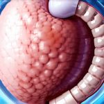
Capsule endoscopy is a non-invasive procedure used to examine the small intestine, an area often difficult to reach with traditional endoscopic methods. It’s becoming increasingly common in diagnosing various gastrointestinal conditions, offering patients a more comfortable alternative to colonoscopies or upper endoscopies for certain issues. This article will delve into how capsule endoscopy works, what conditions it can detect, and what patients can expect before, during, and after the procedure.
Decoding Capsule Endoscopy: A Modern Diagnostic Tool
Capsule endoscopy, also known as video pill or swallowable camera, involves swallowing a small, vitamin-sized capsule containing a miniature camera. This capsule travels naturally through the digestive system, transmitting images wirelessly to a recorder worn externally by the patient. The process allows doctors to visualize areas of the esophagus, stomach, and particularly the small intestine, which is often inaccessible to conventional endoscopy techniques due to its length and tortuous path. Unlike traditional endoscopies that require sedation and can be uncomfortable, capsule endoscopy generally requires minimal preparation and is well-tolerated by most patients. It’s a valuable diagnostic tool for identifying sources of bleeding, inflammation, ulcers, and other abnormalities within the digestive tract, especially when other tests have yielded inconclusive results.
Why it’s Done: Identifying Gastrointestinal Mysteries
Capsule endoscopy is primarily used to investigate unexplained gastrointestinal bleeding, particularly from the small intestine. Traditional methods like colonoscopy and upper endoscopy often miss sources of bleeding in this area, making capsule endoscopy a crucial diagnostic step. It’s also highly effective at detecting Crohn’s disease affecting the small bowel, which can be difficult to visualize with other techniques. Furthermore, it helps identify polyps, tumors (benign or malignant), and ulcers within the digestive tract, offering valuable information for treatment planning.
Specifically, conditions that may require a capsule endoscopy include:
- Unexplained iron deficiency anemia, often indicative of slow, chronic bleeding in the digestive tract.
- Persistent abdominal pain with no clear cause after other investigations.
- Suspected small intestinal tumors or growths.
- Diagnosis and monitoring of Crohn’s disease affecting the small intestine.
- Evaluation of malabsorption syndromes – conditions where nutrients aren’t properly absorbed.
- Identifying sources of recurrent bleeding even after negative colonoscopy and upper endoscopy results.
How to Prepare: Getting Ready for the Procedure
Proper preparation is crucial for a successful capsule endoscopy examination, ensuring clear visualization of the digestive tract. The preparation generally begins with a low-fiber diet several days before the procedure, gradually progressing to a liquid diet 24 hours beforehand. This helps to cleanse the bowel and maximize image quality. Patients are often required to drink a special cleansing solution – a polyethylene glycol (PEG) based solution – the evening before and again on the morning of the test to further clear out the digestive system.
Here’s a typical pre-test checklist:
- Dietary restrictions: Follow your doctor’s instructions regarding low-fiber and liquid diets.
- Medication review: Inform your doctor about all medications you take, including over-the-counter drugs, vitamins, and supplements. Some medications, like iron supplements or blood thinners, may need to be adjusted before the procedure.
- Bowel preparation: Complete the bowel cleansing solution as prescribed by your physician.
- Fasting: Avoid eating or drinking anything for a specified period (usually 8-12 hours) before swallowing the capsule.
- Allergies: Inform your doctor of any allergies you may have, particularly to medications.
What to Expect During the Test: The Process Explained
During the capsule endoscopy procedure itself, the patient simply swallows the capsule with a glass of water. Once swallowed, the capsule begins its journey through the digestive system, transmitting images in real-time to a recorder worn around the waist or attached to a belt. The patient can usually continue their normal activities during this time, but should avoid strenuous exercise and remain within reasonable proximity of the recording device. The process typically takes about 8-12 hours for the capsule to pass through the digestive system and be naturally eliminated during a bowel movement. Patients are instructed to avoid magnetic resonance imaging (MRI) or computed tomography (CT) scans until the capsule has been passed, as these can potentially damage the device.
Understanding the Results: Interpreting What It Means
Once the recording is complete, the images are downloaded and reviewed by a gastroenterologist. The doctor will carefully examine the footage for any abnormalities, such as bleeding points, inflammation, ulcers, or tumors. The results are typically available within a few days to a week, depending on the complexity of the findings and the workload of the interpreting physician. Image quality is crucial, making proper bowel preparation essential for accurate diagnosis.
Is It Safe?: Risks and Side Effects
Capsule endoscopy is generally considered a very safe procedure with minimal risks. However, as with any medical test, there are potential complications to be aware of. The most common side effect is mild abdominal discomfort or cramping during the passage of the capsule. There’s a small risk – less than 1% – that the capsule could become lodged in the digestive tract, potentially requiring surgical intervention for removal. Patients with known strictures (narrowing) or obstructions in their digestive system may not be suitable candidates for this procedure. Rarely, patients might experience nausea or vomiting during the test.
Final Thoughts: A Modern Approach to Digestive Health
Capsule endoscopy provides a valuable and less invasive method for evaluating the small intestine, offering significant benefits for diagnosing various gastrointestinal conditions. It’s particularly useful when traditional endoscopic methods are inconclusive or unable to reach certain areas of the digestive tract. Proper preparation is key to ensuring clear images and accurate results, and while risks are minimal, it’s important to discuss any concerns with your healthcare provider before undergoing the procedure. The technology continues to evolve, offering more detailed insights into the health of our digestive systems.
Questions about this test? Drop them in the comments and we’ll respond.


