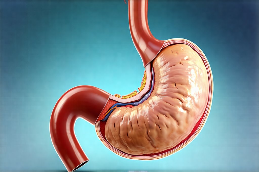
Delayed gastric emptying (DGE), also known as gastroparesis, represents a significant clinical challenge for both patients and healthcare providers. It’s characterized by slower-than-normal passage of food from the stomach into the small intestine. This can lead to a constellation of unpleasant symptoms including nausea, vomiting, bloating, early satiety (feeling full quickly), abdominal pain, and even malnutrition in severe cases. While occasional, mild delays are often benign, persistent DGE substantially impacts quality of life, interfering with daily activities and requiring careful management. Understanding the underlying mechanisms driving delayed emptying is crucial for accurate diagnosis and targeted treatment strategies. However, pinpointing the cause isn’t always straightforward, as various factors – from neurological conditions to medication side effects – can contribute to its development.
The assessment of DGE requires a multifaceted approach, moving beyond simply recognizing symptoms. Traditionally, diagnostic tools have focused on measuring gastric emptying rates, but increasingly sophisticated methods are available to evaluate motility (the movement of the digestive tract), assess potential underlying causes, and even predict treatment response. This article will explore several key tools employed in understanding delayed gastric emptying, outlining their strengths, limitations, and how they contribute to a more comprehensive clinical picture. Importantly, these tools aren’t isolated; they are often used in combination to provide the most accurate assessment of an individual patient’s condition. The goal is not merely diagnosis but also to tailor management strategies for optimal outcomes. Considering practical tools can help with symptom management alongside diagnostic procedures.
Diagnostic Tools Assessing Gastric Emptying Rate
The cornerstone of DGE diagnosis remains assessing how quickly the stomach empties its contents. Historically, this has been achieved through gastric emptying studies, often utilizing solid and liquid meals radiolabeled with isotopes. These studies provide a quantitative measure of gastric emptying half-time – the time it takes for 50% of the ingested material to leave the stomach. A prolonged half-time indicates delayed emptying. However, these tests aren’t without their drawbacks. They require specialized equipment, involve radiation exposure (albeit generally low), and can be affected by factors like patient positioning and meal composition. Newer techniques are attempting to address some of these limitations. Understanding the role of gastric emptying is key in this process.
Beyond traditional scintigraphy (the use of radioactive tracers), breath testing is emerging as a non-radiological alternative. The acetylene breath test leverages the fact that undigested carbohydrates, when reaching the small intestine, are fermented by bacteria producing detectable levels of hydrogen and methane in exhaled breath. A slower rate of gastric emptying leads to more fermentation within the stomach and altered breath gas patterns. While less precise than scintigraphy for quantifying emptying rates, it offers a radiation-free option suitable for certain patient populations. The accuracy of breath tests can also be influenced by factors like intestinal flora composition and recent antibiotic use, requiring careful interpretation.
It’s important to remember that gastric emptying studies provide information about rate, but not necessarily the underlying mechanism causing delay. For example, a slow emptying rate could result from impaired stomach contractility (the ability of the stomach muscles to propel food forward) or dysfunction in the pylorus – the valve controlling the passage of food into the small intestine. Further investigations are often necessary to distinguish between these possibilities and identify the root cause. Gastric emptying studies are valuable but represent only one piece of the puzzle.How gastric emptying rate impacts symptoms should be considered during evaluation.
Motility Studies: Unveiling Digestive Movement
Motility studies delve deeper into the mechanics of digestion, assessing the contractile activity within the stomach and small intestine. High-resolution manometry is a sophisticated technique that utilizes sensors placed within the esophagus or stomach to measure pressure changes associated with muscle contractions. This allows for detailed mapping of peristaltic waves – the coordinated muscular movements that propel food along the digestive tract. In DGE, manometry can reveal abnormalities in gastric contractility, such as weak or uncoordinated contractions, contributing to delayed emptying.
The interpretation of manometric data requires significant expertise and isn’t always straightforward. Normal motility patterns can vary significantly between individuals, making it challenging to establish definitive thresholds for abnormality. However, manometry is particularly useful in differentiating between different types of gastroparesis – for example, identifying cases caused by impaired contractility versus those primarily related to pyloric dysfunction. Wireless capsule motility studies offer another approach, using ingestible sensors to monitor transit time and pressure changes throughout the gastrointestinal tract. This provides a less invasive alternative to traditional manometry but may not provide the same level of detail.
Understanding the interplay between gastric and intestinal motility is vital. DGE isn’t solely about how slowly food leaves the stomach; it’s also influenced by how effectively the small intestine receives and processes that food. Motility studies offer a more nuanced understanding of digestive function than emptying rate alone. These tests help physicians pinpoint specific areas of dysfunction and guide treatment decisions. Options for monitoring slow motility are also available.
Imaging Techniques: Visualizing Structural and Functional Issues
While functional assessments like motility studies are crucial, imaging techniques provide valuable structural information and can identify potential underlying causes of DGE. Upper endoscopy allows direct visualization of the stomach and duodenum (the first part of the small intestine), enabling detection of structural abnormalities such as ulcers, inflammation, or obstructions that might contribute to delayed emptying. It also permits biopsy sampling for microscopic examination, helping to rule out conditions like gastritis or celiac disease.
Beyond traditional endoscopy, capsule endoscopy offers a non-invasive way to visualize the small intestine. A tiny camera encased in a swallowable capsule transmits images as it travels through the digestive tract, providing a more comprehensive assessment of intestinal health. This is particularly useful for identifying subtle abnormalities that might be missed during upper endoscopy. However, capsule endoscopy doesn’t allow for biopsy sampling or intervention.
Finally, magnetic resonance imaging (MRI) and computed tomography (CT) scans can provide detailed anatomical images of the abdomen, revealing structural anomalies like tumors or masses that could be compressing the stomach or duodenum. Functional MRI techniques are also emerging, offering the potential to assess gastric motility in vivo without requiring invasive procedures. Imaging techniques complement functional assessments by providing a broader understanding of the underlying pathology. They help identify both the cause and the consequence of delayed emptying, guiding diagnostic and treatment decisions. Patients may also benefit from evaluating tools for identifying silent inflammation.
It’s crucial to acknowledge that diagnosing DGE isn’t always easy. Many patients present with nonspecific symptoms, making it challenging to differentiate gastroparesis from other conditions like functional dyspepsia (indigestion). A combination of these tools – gastric emptying studies, motility assessments, and imaging techniques – is often necessary to arrive at an accurate diagnosis and develop a tailored treatment plan that addresses the specific needs of each patient. Furthermore, ongoing research continues to refine existing diagnostic methods and explore new technologies for better understanding and managing this complex condition. Modern tools for monitoring acid reflux can also assist in a comprehensive evaluation.
Dr. Olivia Bennett, MD is a board-certified gastroenterologist specializing in advanced digestive diagnostics. With over 12 years of clinical experience, she focuses on non-invasive testing methods, gut microbiome assessment, and functional GI disorders. Dr. Bennett is passionate about helping patients understand their digestive health through clear and accessible medical insights.