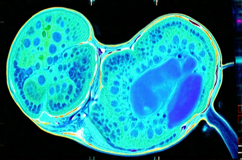
Gallbladder issues are surprisingly common, often sneaking up on individuals with vague symptoms that can easily be attributed to other causes. This makes early detection crucial for preventing more serious complications like gallstones, cholecystitis (inflammation of the gallbladder), and even pancreatic involvement. While many people associate gallbladder problems with sudden, intense pain, a significant number experience subtle signs that warrant investigation. Recognizing these initial indicators and utilizing appropriate diagnostic tools allows for timely intervention and management, often avoiding the need for extensive treatment or surgery. The good news is medical imaging has advanced considerably, providing non-invasive methods to assess gallbladder health even before symptoms become debilitating.
The gallbladder, a small organ nestled under the liver, plays a vital role in digestion. It stores bile – a fluid produced by the liver that helps break down fats. When we eat fatty foods, the gallbladder releases this bile into the small intestine. Problems arise when the flow of bile is disrupted, leading to stone formation or inflammation. The early stages often present with nonspecific symptoms like bloating, indigestion, or mild discomfort in the upper right abdomen. It’s important to remember that these symptoms can mimic other conditions, highlighting the necessity of accurate diagnostic testing. This article will explore the various digestive scans available for detecting early gallbladder trouble, focusing on their strengths and limitations.
Imaging Techniques for Gallbladder Assessment
Several imaging techniques are routinely employed to visualize the gallbladder and identify potential problems. The choice of scan depends on the suspected issue, patient history, and accessibility. Ultrasound is often the first-line investigation due to its affordability, non-invasiveness, and lack of ionizing radiation. However, other modalities like CT scans, MRI, and HIDA scans offer more detailed information or are used when ultrasound findings are inconclusive. Early detection relies on utilizing these tools proactively, especially for individuals with risk factors such as family history of gallbladder disease, obesity, rapid weight loss, or certain medical conditions. It’s important to discuss your personal risks with a healthcare professional to determine the most appropriate screening approach. You might also consider looking into home kits that can detect digestive infections as part of preventative care.
Ultrasound uses sound waves to create images of internal organs. It’s particularly effective at detecting gallstones – the most common cause of gallbladder problems. A technician will apply a gel to your abdomen and then move a transducer over the skin, sending out sound waves that bounce back differently depending on the tissue they encounter. These echoes are converted into an image displayed on a screen. While excellent for visualizing stones, ultrasound can sometimes struggle with patients who have significant body habitus or gas in the intestines, potentially obscuring the gallbladder. Understanding initial scans used before more invasive digestive tests is crucial for a proper diagnosis.
CT (Computed Tomography) scans and MRI (Magnetic Resonance Imaging) offer more detailed anatomical images than ultrasound. CT scans use X-rays to create cross-sectional images, while MRIs utilize magnetic fields and radio waves. While both are excellent for identifying structural abnormalities, they involve radiation (CT) or longer scan times and potential claustrophobia (MRI). They’re typically reserved for cases where ultrasound is inconclusive or when more information about surrounding structures is needed. MRI specifically excels at visualizing the biliary ducts – the tubes that carry bile from the gallbladder to the small intestine.
Understanding Specific Scans in Detail
Beyond the standard imaging techniques, specialized scans provide even greater insight into gallbladder function and potential blockages. The HIDA scan (hepatobiliary iminodiacetic acid scan), also known as a cholescintigraphy scan, is particularly useful for assessing gallbladder emptying and identifying obstructions. This scan involves intravenously injecting a small amount of radioactive tracer that is then taken up by the liver and excreted into the bile. A gamma camera tracks the movement of the tracer, allowing doctors to visualize the flow of bile from the liver, through the biliary ducts, and eventually into the gallbladder and small intestine.
The HIDA scan can detect acute cholecystitis – inflammation of the gallbladder – even when ultrasound findings are normal. If the tracer doesn’t reach the gallbladder within a specified timeframe (typically 4-6 hours), it suggests an obstruction, often due to gallstones or inflammation. Another specialized technique is MRCP (Magnetic Resonance Cholangiopancreatography), which uses MRI to visualize the biliary ducts in great detail without requiring invasive procedures like endoscopic retrograde cholangiopancreatography (ERCP). MRCP is becoming increasingly popular for diagnosing bile duct stones, strictures, and other abnormalities. If you’re experiencing digestive issues alongside neurological symptoms, tests that connect digestive and neurological symptoms might be helpful in finding the root cause.
Detecting Early Signs with Ultrasound
Ultrasound remains a cornerstone of initial gallbladder assessment due to its accessibility and cost-effectiveness. However, interpreting ultrasound images requires expertise, as subtle findings can indicate early trouble. – A key indicator is the presence of sludge within the gallbladder – a precursor to stone formation. Sludge appears as a cloudy or hazy substance on the ultrasound image. – Another sign to watch for is thickening of the gallbladder wall, which suggests inflammation. – Furthermore, even small gallstones may be identified early with high-resolution ultrasound technology.
It’s important to note that not all gallstones cause symptoms. Many individuals have asymptomatic gallstones – stones that are present but don’t produce any noticeable problems. These “silent” stones can still lead to complications down the line if left untreated, making routine screening beneficial for at-risk individuals. The effectiveness of ultrasound is enhanced when performed by a skilled sonographer and interpreted by an experienced radiologist or physician. Periodic follow-up ultrasounds may be recommended to monitor changes in gallbladder status. It’s also worth understanding why standard scans might miss early gut problems.
The Role of HIDA Scans in Functional Assessment
HIDA scans offer valuable functional information that complements structural imaging like ultrasound and CT scans. Unlike these modalities, which primarily show the anatomy, HIDA scans assess how well the gallbladder is working – specifically, its ability to contract and empty bile. This scan is particularly useful for diagnosing acalculous cholecystitis – inflammation of the gallbladder in the absence of stones.
The process typically involves: 1) Intravenous injection of the radioactive tracer. 2) Imaging with a gamma camera over several hours. 3) Assessment of tracer uptake by the liver and excretion into the biliary system. A normal HIDA scan shows rapid filling of the gallbladder, followed by contraction in response to stimulation (often with CCK – cholecystokinin). Failure of the gallbladder to fill or contract suggests a problem.
Interpreting Scan Results & Next Steps
Interpreting digestive scans requires expertise and should always be done by qualified medical professionals. A positive scan result doesn’t automatically mean surgery is necessary. The treatment plan depends on the specific findings, symptom severity, and overall health of the patient. – Mild sludge without symptoms may require only observation and lifestyle modifications (e.g., a low-fat diet). – Gallstones causing intermittent pain might be managed with medication to dissolve the stones or watchful waiting. – Severe inflammation or obstruction often necessitates gallbladder removal – typically via laparoscopic cholecystectomy, a minimally invasive surgical procedure.
It’s crucial to discuss scan results thoroughly with your doctor and ask questions about all available treatment options. Don’t hesitate to seek a second opinion if you have concerns. Proactive screening and early detection are essential for preventing complications and maintaining optimal gallbladder health. Remember that imaging scans are just one piece of the puzzle – a comprehensive evaluation includes a detailed medical history, physical examination, and potentially blood tests to assess liver function. Scans and labs that help detect gallbladder dysfunction can provide crucial insights for personalized care. Furthermore, understanding how digestive tests help explain low appetite and early satiety can aid in holistic assessment. If surgery is considered, digestive tests that should be done before surgery are vital for preparation and planning.


