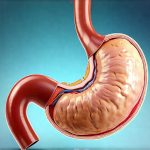
Gastric emptying scintigraphy is a non-invasive nuclear medicine test used to assess how quickly food empties from the stomach into the small intestine. It’s a valuable diagnostic tool for individuals experiencing symptoms related to delayed gastric emptying, often referred to as gastroparesis, or rapid gastric emptying. This article will provide a comprehensive overview of this procedure, covering its purpose, preparation, execution, interpretation, and potential risks, empowering you with knowledge about what to expect if your doctor recommends it.
Unveiling Gastric Emptying Scintigraphy: A Detailed Look
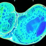
Gastric emptying scintigraphy utilizes a small amount of radioactive tracer mixed with a standard meal to visually track the rate at which food leaves the stomach. Unlike more invasive methods like endoscopy or biopsies, this test provides functional information about gastric motility – how well the stomach muscles work to propel food along the digestive tract. The process involves ingesting the prepared meal and then having images taken over several hours using a gamma camera, which detects the radiation emitted by the tracer. These images create a dynamic visualization of the emptying process, allowing clinicians to identify abnormalities in gastric transit time. This allows for precise assessment, leading to more targeted treatment strategies for patients experiencing digestive issues.
Why It’s Done: Identifying and Diagnosing Gastric Motility Disorders
This test is primarily employed to diagnose and evaluate conditions affecting gastric emptying rates. Delayed gastric emptying (gastroparesis) can cause a range of uncomfortable symptoms including nausea, vomiting, bloating, early satiety (feeling full quickly), and abdominal pain. The scintigraphy helps determine the severity of the delay, aiding in differentiating between various causes of gastroparesis. These causes can include diabetes mellitus, post-surgical changes, certain medications, or idiopathic (unknown) reasons. Conversely, rapid gastric emptying – though less common – can lead to symptoms like diarrhea and dumping syndrome, where food moves too quickly into the small intestine, causing osmotic imbalances.
Beyond diagnosing gastroparesis, this test is also valuable in:
* Monitoring the effectiveness of treatments for gastroparesis.
* Evaluating patients before considering surgical interventions, such as gastric pacemaker implantation.
* Identifying underlying causes of chronic digestive symptoms when other tests are inconclusive.
* Assessing nutritional status in patients with suspected malabsorption issues related to altered gastric emptying.
How to Prepare: Getting Ready for the Procedure
Proper preparation is crucial for accurate results. Your doctor will provide specific instructions tailored to your individual needs, but generally include these steps:
- Fasting: You’ll likely need to fast for at least six hours before the test. This ensures an empty stomach for optimal imaging and minimizes interference with tracer uptake.
- Medication Review: Inform your doctor about all medications you are taking, including over-the-counter drugs and supplements. Certain medications can affect gastric emptying and may need to be temporarily adjusted or discontinued prior to testing. Specifically, medications that slow down the digestive system like opioids should be discussed with your physician.
- Dietary Restrictions: Avoid heavy meals on the day before the test. A light dinner is usually recommended.
- Hydration: Staying adequately hydrated is important, so drink clear fluids as instructed by your doctor.
- Pregnancy Notification: If you are pregnant or suspect you might be, inform your doctor immediately. Nuclear medicine tests involve radiation exposure and precautions must be taken.
What to Expect During the Test: A Step-by-Step Guide
The gastric emptying scintigraphy procedure typically takes 2-4 hours to complete, depending on the individual’s emptying rate and the specific protocol used by the facility. It begins with the administration of the radioactive tracer, usually Technetium-99m (Tc-99m), mixed into a standard meal – often eggs or oatmeal blended with water. The amount of radioactivity is very low and considered safe for most individuals.
- Tracer Ingestion: You will ingest the prepared meal containing the tracer.
- Initial Imaging: A baseline image is taken immediately after ingestion to establish the initial distribution of the tracer in the stomach.
- Serial Imaging: Images are then acquired at regular intervals (e.g., 30 minutes, 1 hour, 2 hours, and potentially longer) using a gamma camera positioned over your abdomen. The gamma camera detects the radiation emitted by the tracer as it moves from the stomach into the small intestine.
- Positioning: During imaging, you will likely be asked to remain relatively still, although some facilities allow for limited movement between images. You may be seated or lying down depending on the equipment and facility protocols.
- Hydration: You’ll usually be encouraged to sip water throughout the procedure to stay hydrated.
Understanding the Results: Interpreting What It Means
The results are interpreted by a nuclear medicine physician who analyzes the images and calculates several key parameters, including:
- Half-Emptying Time (T1/2): The time it takes for half of the ingested meal to empty from the stomach.
- Total Emptying Rate: The percentage of the meal that has emptied after a specific period.
- Gastric Retention at Specific Time Points: Amount of radioactive tracer remaining in the stomach at certain intervals.
Normal values vary slightly between laboratories and protocols, but generally:
- A half-emptying time of over 60-90 minutes suggests delayed gastric emptying (gastroparesis).
- A rapid half-emptying time (less than 30-45 minutes) may indicate rapid gastric emptying.
These values are then correlated with your symptoms and other clinical findings to make an accurate diagnosis. Factors such as diabetes, medication use, and previous surgeries will also be considered during interpretation.
Is It Safe? Risks and Side Effects
Gastric emptying scintigraphy is generally considered safe, but like any medical procedure, it carries minimal risks:
- Radiation Exposure: The amount of radiation exposure from this test is low – comparable to that experienced during a cross-country flight or a routine X-ray.
- Allergic Reaction: Although rare, some individuals may experience mild allergic reactions to the tracer.
- Discomfort: Some patients might experience mild discomfort from lying still for an extended period.
- Nausea/Vomiting: Occasionally, nausea or vomiting may occur during the test, particularly if you have existing digestive issues.
Pregnant women should avoid this test due to the risk of radiation exposure to the fetus. Individuals with kidney problems should inform their doctor as they may require adjustments to ensure proper tracer elimination.
Final Thoughts: A Comprehensive Assessment Tool
Gastric emptying scintigraphy offers a valuable, non-invasive way to assess gastric function and diagnose motility disorders that can cause significant digestive distress. By providing objective data on the rate at which food empties from the stomach, it helps clinicians pinpoint the underlying causes of symptoms and tailor treatment plans accordingly. This test is an important part of evaluating patients with suspected gastroparesis or other conditions affecting digestion.
Questions about this procedure? Drop them in the comments below and we’ll respond as soon as possible!


